High Back Muscles Diagram / Oversized Muscle Identification Diagrams!- Great for ... : Attached to the bones of the skeletal system are about 700 named muscles that make up roughly half of a person's body weight.
High Back Muscles Diagram / Oversized Muscle Identification Diagrams!- Great for ... : Attached to the bones of the skeletal system are about 700 named muscles that make up roughly half of a person's body weight.. Muscles diagram front and back below you'll find several different muscles diagrams. The superficial back muscles are covered by skin, subcutaneous connective tissue and a layer of fat. How to build a wide back. The diagram is a common one used to explain sliding filament theory, but don't worry about trying to gluteus maximus, biceps femoris, semitendinosus, semimembranosus at the back and the adductor or groin major muscles of the neck and back include the erector spinae, multifidus, rectus abdominus. Start studying back muscle diagrams.
Start with the anatomy of the deep muscles of the back by exploring our videos, quizzes, labeled diagrams, and articles. Other muscles are small and cover much less space. High quality images of interesting designs, including architectural, graphic, industrial, furniture & product design. The following diagram shows all the major back muscles. Luckily you've found this page to help you.

Skeletal muscles are the only muscles that can be consciously controlled.
This muscle diagram made to look like a human. Human muscle system, the muscles of the human body that work the skeletal system, that are under voluntary control, and that are concerned with movement, posture, and balance. Within this group of back muscles you will find the latissimus dorsi, the trapezius, levator scapulae and the the intrinsic (deep) muscles of the backcan be further subdivided into their own superficial, intermediate and deep layers. Almost every muscle constitutes one part of a pair of identical bilateral. There are anterior muscles diagrams and posterior. In all its forms, it makes up nearly half of the body's mass. Now label the diagram in your workbook! The superficial back muscles are covered by skin, subcutaneous connective tissue and a layer of fat. Equally important is the erector spinae muscles. Short of a great deal of descriptive text, the easiest way to answer this is with illustrations. Their main function is contractibility. This image is titled back muscles diagram and is attached to our article about best back muscles training exercises. The back comprises the dorsal part of the neck and the torso (dorsal body cavity) from the occipital bone to the top of the tailbone.
When we think of back muscles, latissimus dorsi (lats) comes to mind. Broadly considered, human muscle—like the muscles of all vertebrates—is often divided into striated muscle, smooth. This is a table of skeletal muscles of the human anatomy. Any exercise you do specifically for shaping your back is definitely very visible and noticeable, and one of the most important things to making your whole body look more muscular. Muscles of the back can be divided into superficial, intermediate, and deep group.since the all the back muscles originate in embryo (fetus) form by locations other than the back, muscles in the.
The back comprises the dorsal part of the neck and the torso (dorsal body cavity) from the occipital bone to the top of the tailbone.
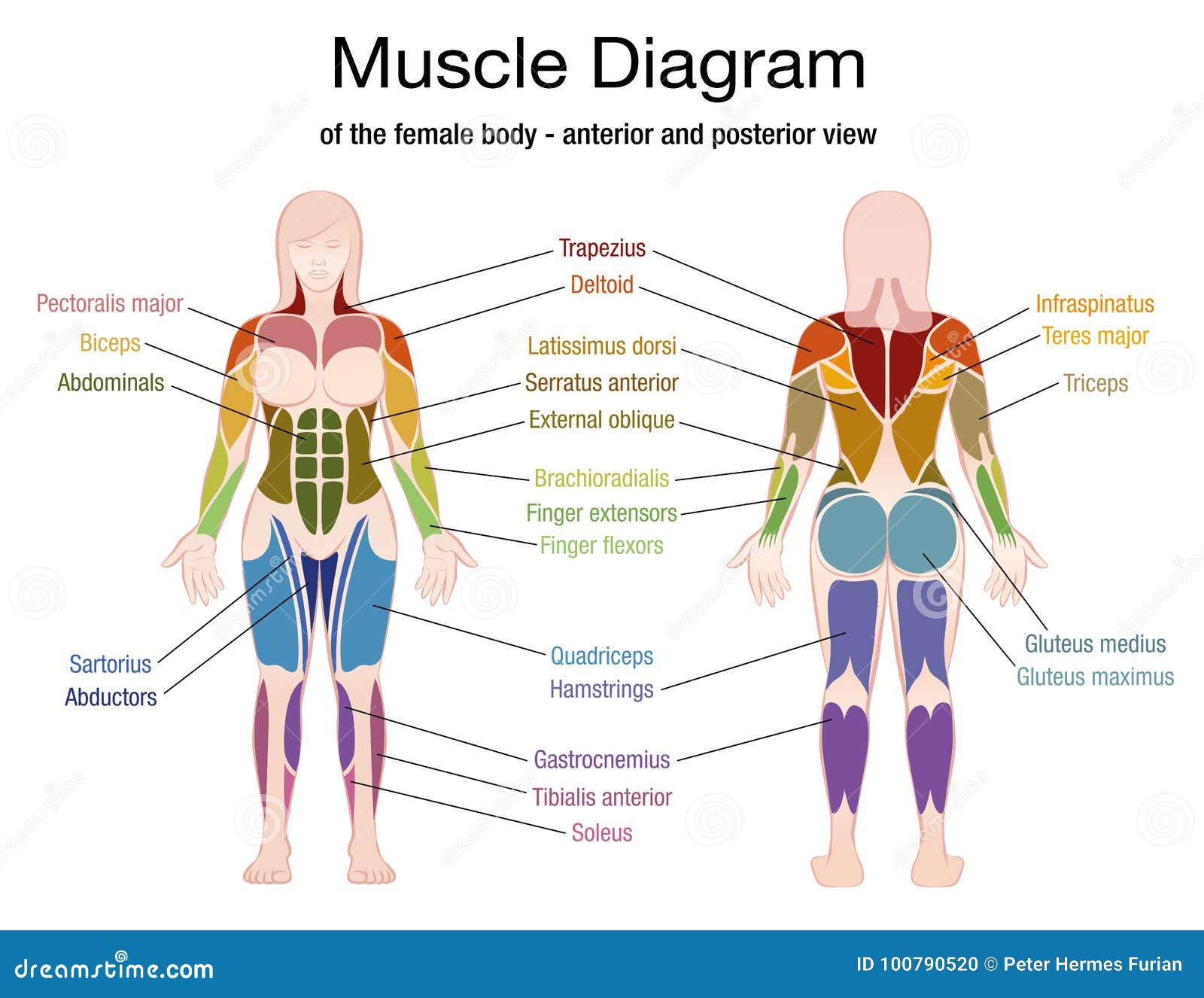
The superficial back muscles are the muscles found just under the skin. Start with the anatomy of the deep muscles of the back by exploring our videos, quizzes, labeled diagrams, and articles. The diagram is a common one used to explain sliding filament theory, but don't worry about trying to gluteus maximus, biceps femoris, semitendinosus, semimembranosus at the back and the adductor or groin major muscles of the neck and back include the erector spinae, multifidus, rectus abdominus. But muscle is also the dominant tissue in the heart and in the walls of other hollow organs of the body. All of these things can lead to long term back pain (and chronic complaining!). Their main function is contractibility. Attached to the bones of the skeletal system are about 700 named muscles that make up roughly half of a person's body weight. This is a table of skeletal muscles of the human anatomy. Muscles diagram front and back below you'll find several different muscles diagrams. Skeletal muscles are the only muscles that can be consciously controlled. Other muscles are small and cover much less space. Flexibility tips muscle diagram hamstring muscles body chart latissimus dorsi tight hamstrings. Luckily you've found this page to help you.
20 turkish airlines flight 981 photos and premium high res pictures. The back has some of the body's largest muscles (erector spinae group) and some of the smallest and most numerous ones. Equally important is the erector spinae muscles. There are around 650 skeletal muscles within the typical human body. They are attached to bones, and muscles in the torso protect the internal organs at the front, sides, and back of the body.

Most will label a diagram of muscle with its structures.
High quality images of interesting designs, including architectural, graphic, industrial, furniture & product design. Start studying back muscle diagrams. Short of a great deal of descriptive text, the easiest way to answer this is with illustrations. The muscles of the back can be divided in three main groups according to their anatomical position and function. The back comprises the dorsal part of the neck and the torso (dorsal body cavity) from the occipital bone to the top of the tailbone. The back's muscles start at the top of the back (named the cervical vertebrae) and go to the tailbone (also named the coccyx). The diagram is a common one used to explain sliding filament theory, but don't worry about trying to gluteus maximus, biceps femoris, semitendinosus, semimembranosus at the back and the adductor or groin major muscles of the neck and back include the erector spinae, multifidus, rectus abdominus. Their main function is contractibility. But muscle is also the dominant tissue in the heart and in the walls of other hollow organs of the body. This is a table of skeletal muscles of the human anatomy. Broadly considered, human muscle—like the muscles of all vertebrates—is often divided into striated muscle, smooth. Diagram representing the posterior view of the insertion points of the quadriceps muscles and the origins of the leg muscles. Other muscles are small and cover much less space.


Komentar
Posting Komentar